Our research group is located in the Physics Department and Center for Computational and Integrative Biology (CCIB) at Rutgers University - Camden. We use high performance computing and Molecular Dynamics Simulation to investigate biophysical interactions of proteins, membranes, and small molecules. Many of our research topics involve using concepts and methods from physics to understand complex signaling mechanisms in the central nervous system. Our group members come from a range of backgrounds, including biology, pharmacology, physical chemistry, engineering and physics.
FEATURED LAB ART
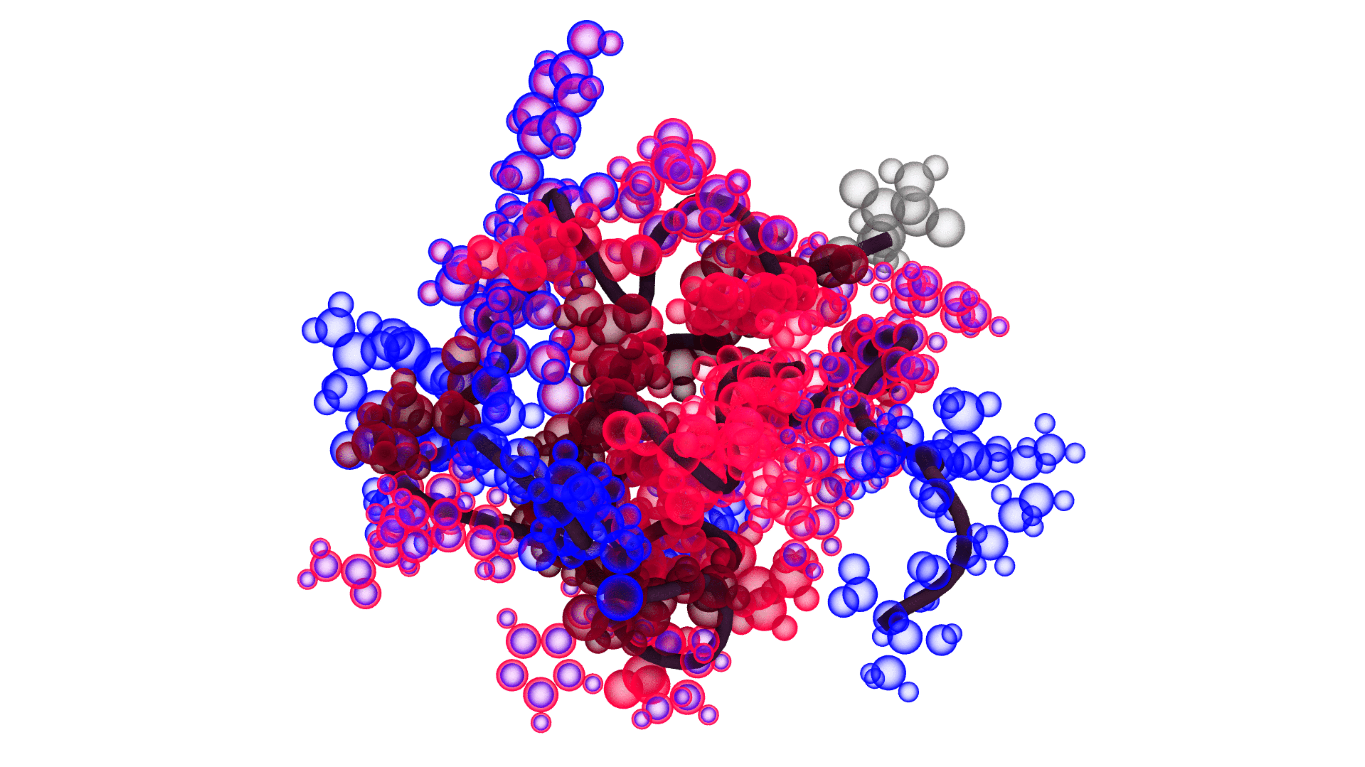
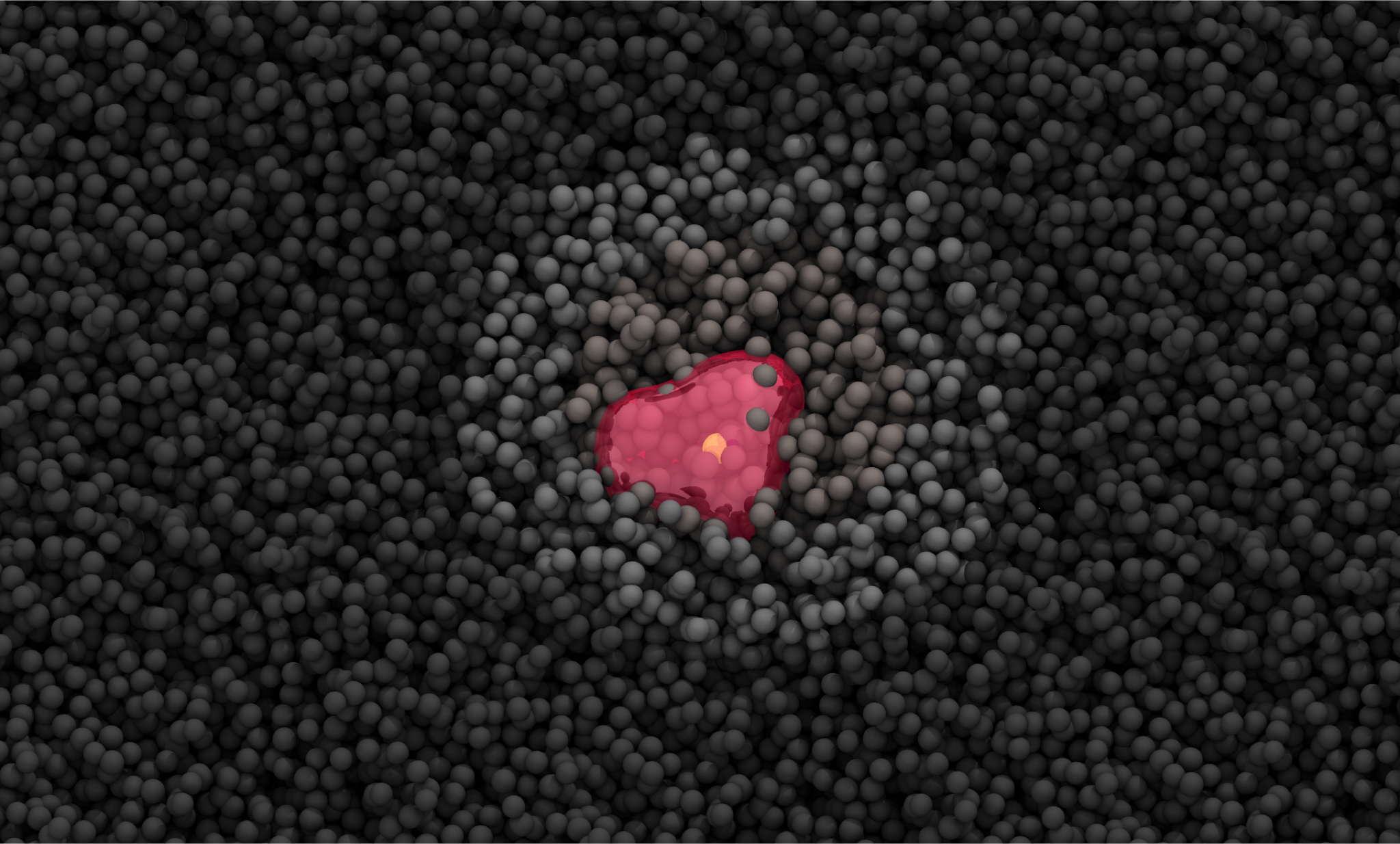
In a bind
The average configuration of ELIC (orange surface) with a tightly bound POPG (gray spheres). The radius of each sphere of POPG is inversely proportional to the standard deviation of its coordinates. Data were collected from the last of a simulation. This binding mode is stabilized in part by long-lived hydrogen bonds to the phosphate and glycerol backbone of the lipid (largest spheres). Image created in VMD with post-processing in Photoshop.
VMD IMAGE CONTEST WINNER 2025
Image Credit: Ezry Santiago-McRae
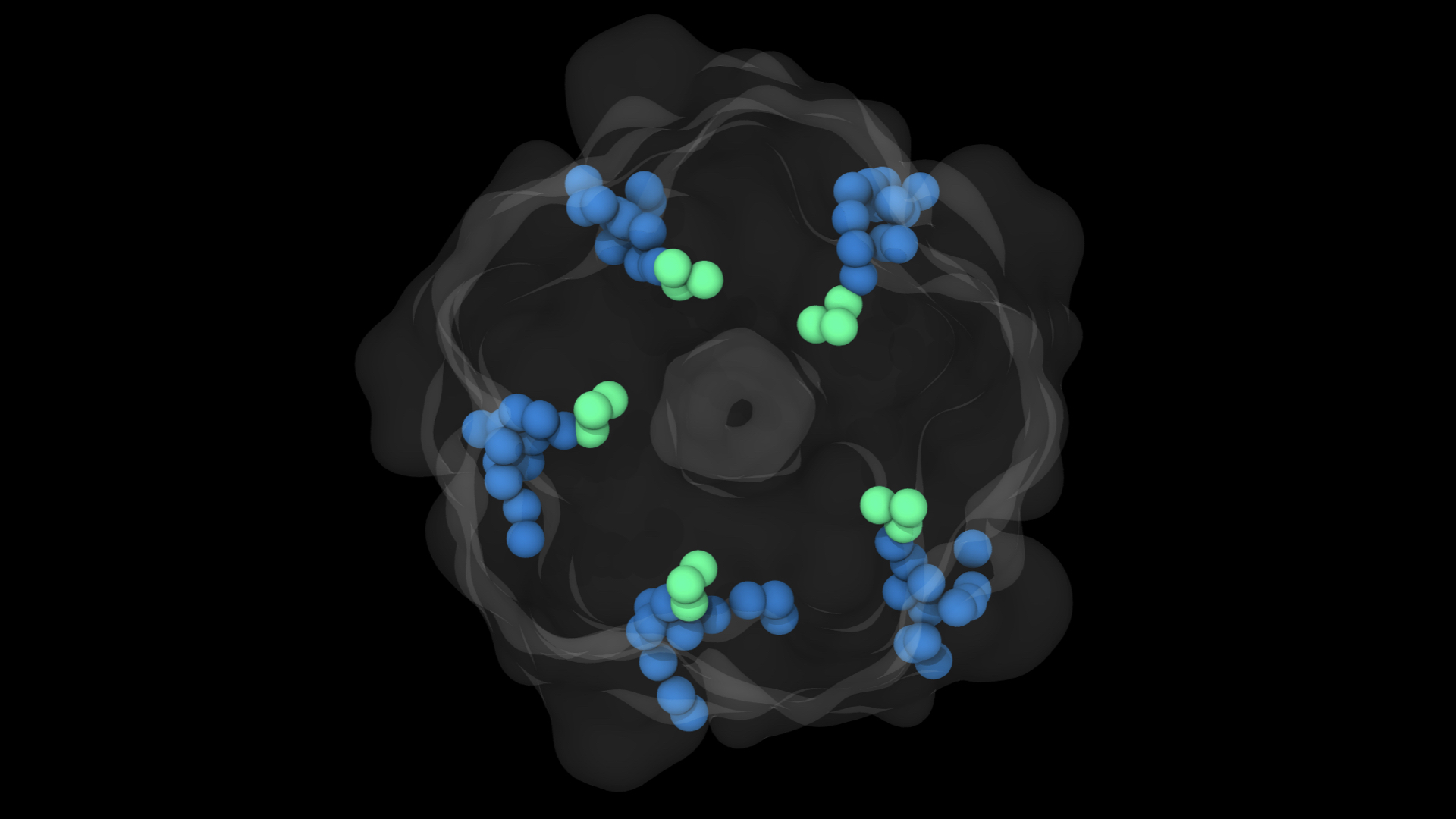
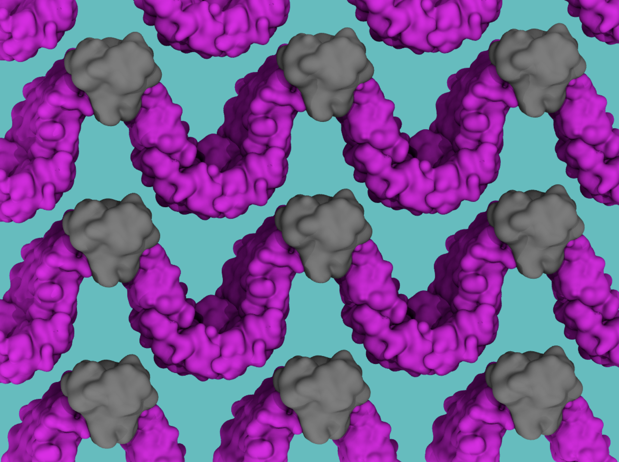
Can you find the binding lipid?
POPG molecules in a 2:1:1 POPC:POPE POPG membrane diffuse around and interact with ELIC5, an ‘open state` 5-mutant of the ELIC pentameric Ligand-Gated Ion Channel. Several binding events occur, but one stands out. Can you find the binding lipid?
VMD MOVIE CONTEST WINNER 2025
Image Credit: Jesse Sandberg
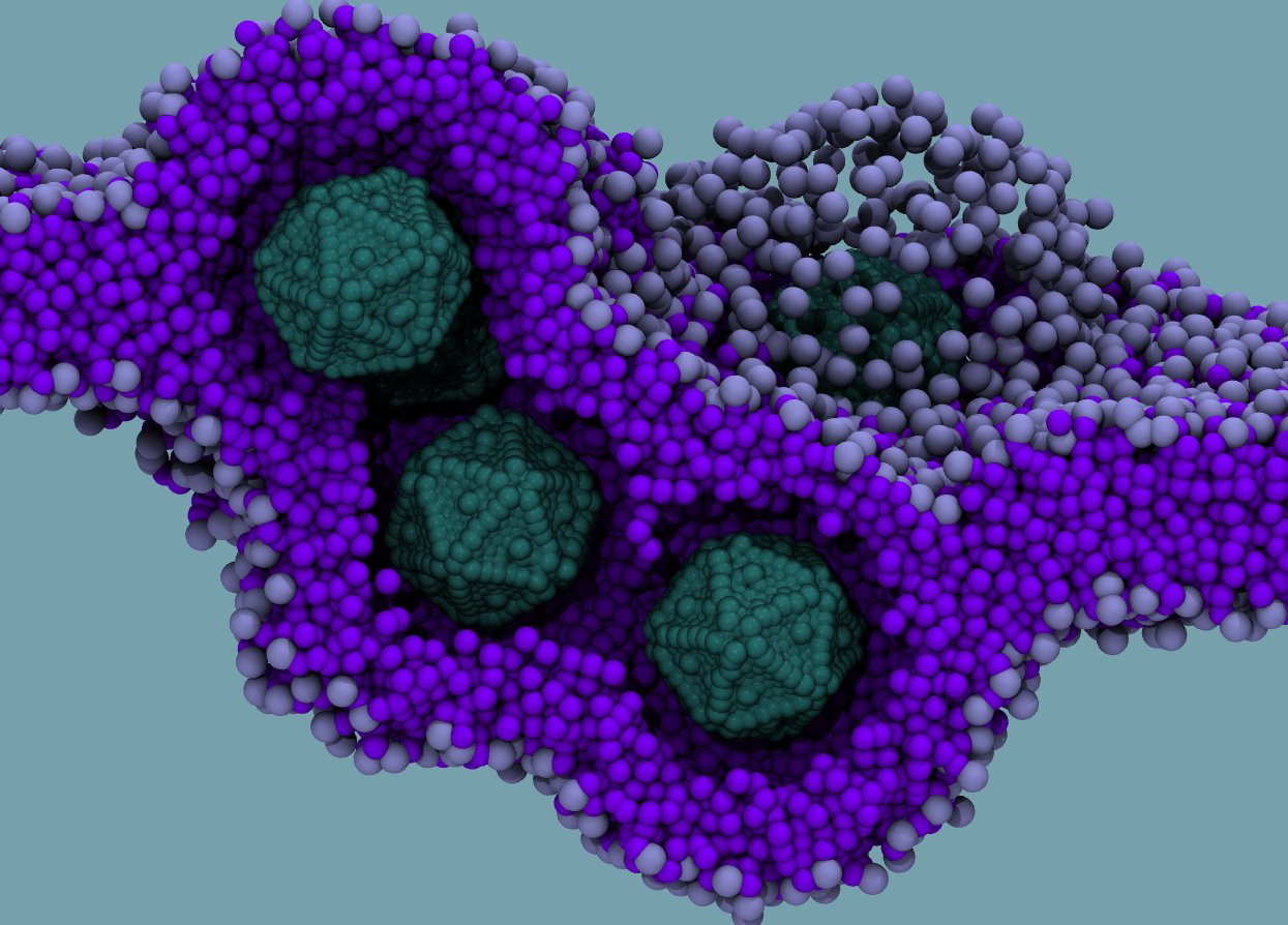
Positive Infinity
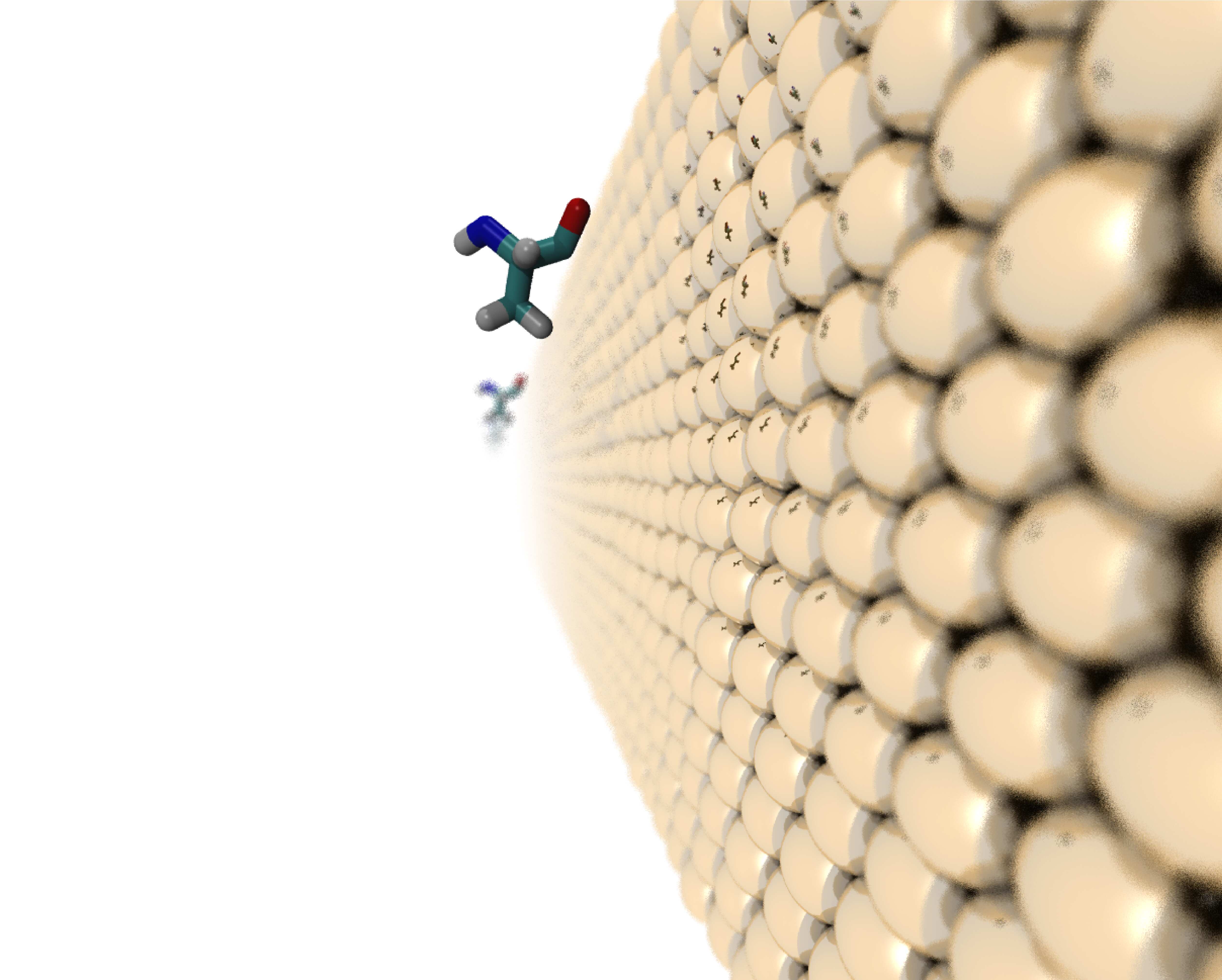
Scientific Impact: In ABF a reaction coordinate is defined, then using a thermodynamic integration like approach, the free energy along that reaction coordinate is measured. Here I am showing an infinite gold slab and an alanine being pulled away from it along the z direction. We use the free energy profiles to calculate the adsorption free energy of alanine to the gold surface. The adsorption free energies of these atomistic systems are used to determine better coarse-grained models of gold. Artist Concept: “The mechanism of eqn. (8.2.1) is one in which an imaginary external controlling influence (‘hand of God’ in the form of the lambda parameter)…” - Tuckerman. Here I play with the idea of infinity and how things that appear finite may in fact be infinite.
VMD IMAGE CONTEST RUNNER UP 2025
Image Credit: Jahmal Ennis
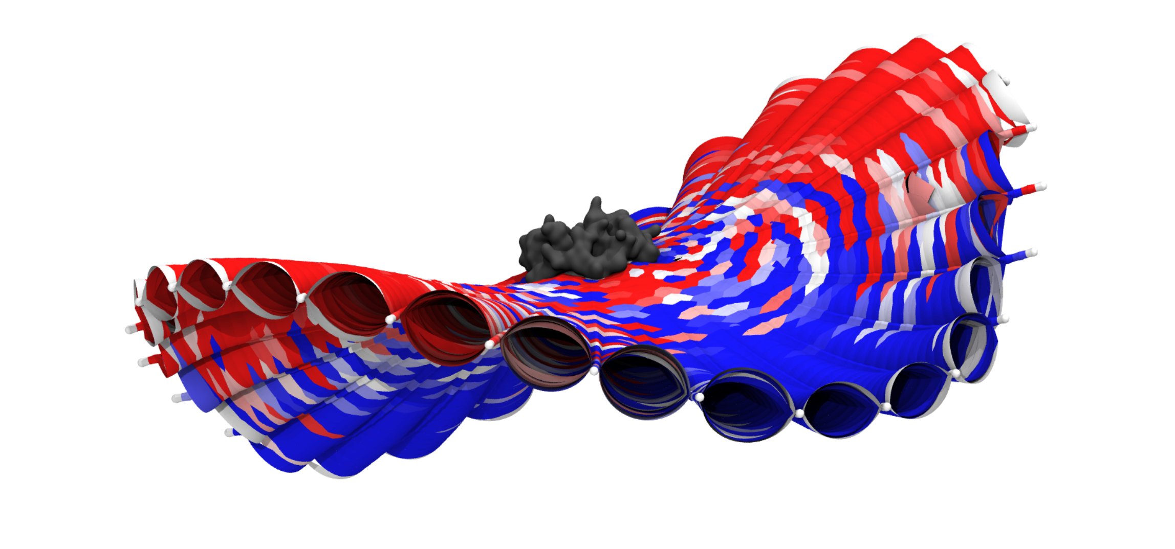
Ghostly gathering
The inositol head group (green) of PAPI inserts into the intersubunit cleft of the a-7 nicotinic acetylcholine receptor N214F mutant (translucent grey) when simulated in an oocyte-mimetic membrane background.
VMD IMAGE CONTEST RUNNER UP 2025
Image Credit: Jesse Sandberg
Looking glass
Temperature replica exchange molecular dynamics (T-REMD) allows a protein to explore many conformations as it traverses rungs of a temperature “ladder”. This movie shows a Beta Amyloid A21M mutant from a T-REMD simulation at 5 different temperatures (low to high: purple, blue, green, yellow, red).
VMD MOVIE CONTEST RUNNER UP 2025
Movie Credit:Connor Pitman
Sugar and Fat for the brain
When simulated in an oocyte-mimetic membrane, PAPI (olive green) lipids insert their inositol head-groups into the intersubunit cleft of the a-7 nicotinicacetylcholine receptor (grey). Mutating one protein residue in the cleft, N214F, causes increased binding of PAPI when DHA is in the bulk (shown in this video). This effect is not seen when Arachidonic Acid, a similar fatty acid to DHA, is in the bulk.
VMD MOVIE CONTEST RUNNER UP 2025
Movie Credit: Jesse Sandberg
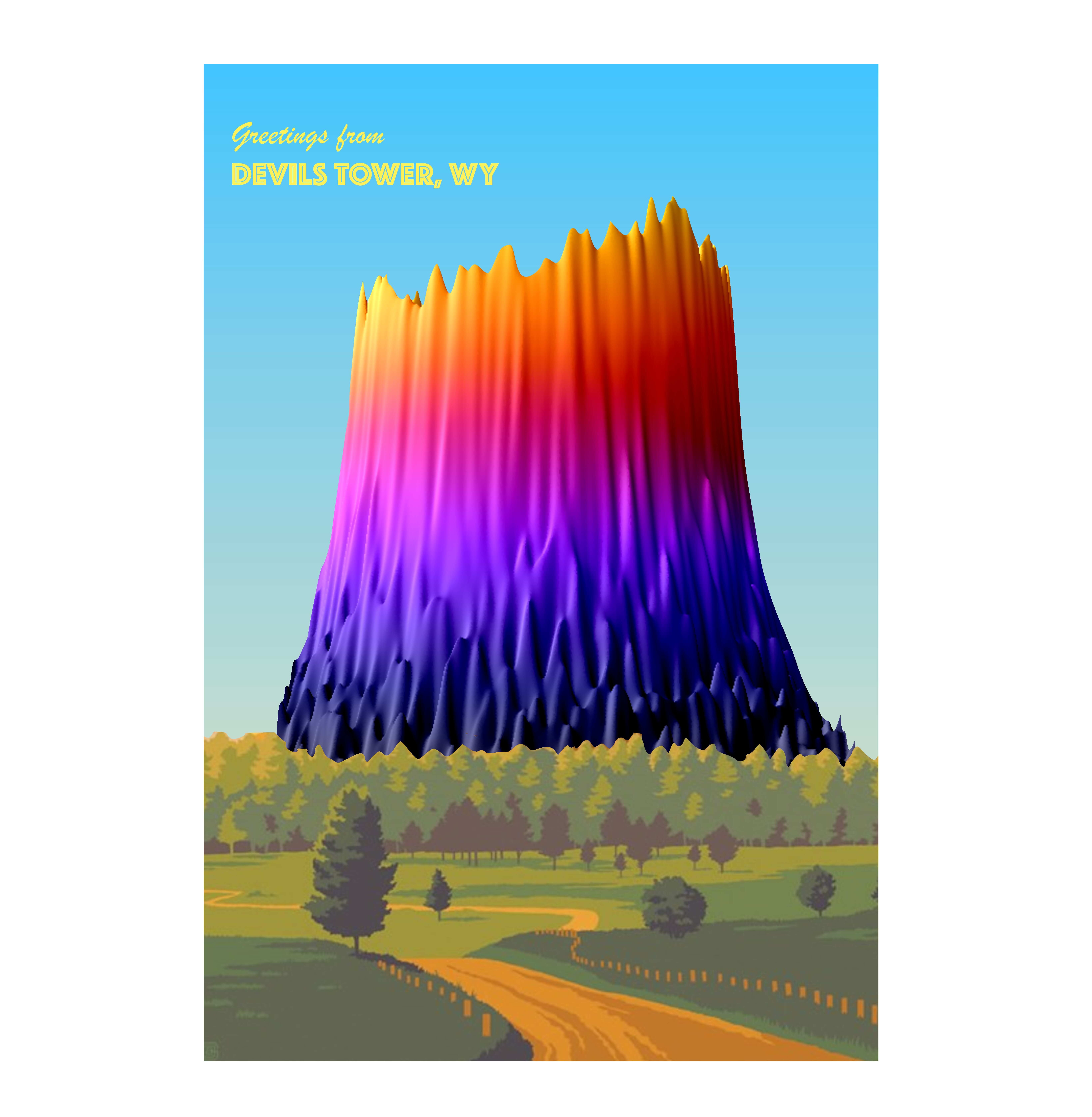
A postcard from Wyoming
A vesicle containing 90% POPC and 10% Ceramide Phosphoglycerate (CPG) is captured via fluorescence microscopy. The intensities in the 2D image have been transformed into a 3D surface plot via Fiji. When viewed from the side, this surface plot looks strikingly similar to the Devils Tower National Monument in Wyoming (popularized in the movie Close Encounters of the Third Kind).
SCIENTIFIC MEDIA CONTEST WINNER 2025
Image Credit: Jesse Sandberg
Under The Surface
A tribute to the blobulator algorithm using the Unity engine. The blobulator wording is made exclusively from simulated synthetic peptides. While some of the wording has multiple peptides blended together, all materials used to form the word came entirely from Gromacs simulations viewed and graphically represented in VMD. The proteins orbiting the text represent each member of the lab: Insulin (Connor) , Amyloid-Beta (Lindsey), BamA (Jahmal), ELIC, (Ezry), Heme (Asim), and nAChR (Jesse). These proteins fluctuate from the blobulated representation to the secondary structure representation when in and out of water, respectively. Whether a protein is intrinsically disordered, globular, or membrane-bound, under the surface, they all rely on hydrophobicity to maintain function.
SCIENTIFIC MEDIA CONTEST WINNER 2025
Movie Credit: Ryan Lamb
Crab!
This image shows a blobulated human tankyrase dimer. Blobs are colored as follows- h-blobs: red, p-blobs: pink, s-blobs: white. The residue Histidine 162 is shown in VDW and colored in white and black.
Image Credit: Connor Pitman
Hydrophobic zipper
Hydrophobic regions, or h-blobs, in the BDNF prodomain sequence act like the teeth of a zipper, transiently interlocking to fasten its termini together. Three prodomain variants, represented in gold, silver, or rose gold, display a zipped state (translucent h-blobs) and an unzipped state (opaque h-blobs) in their conformational ensemble. Each polyhedron in the zipped state represents a hydrophobic amino acid.
Image Credit: Lindsey Riggs
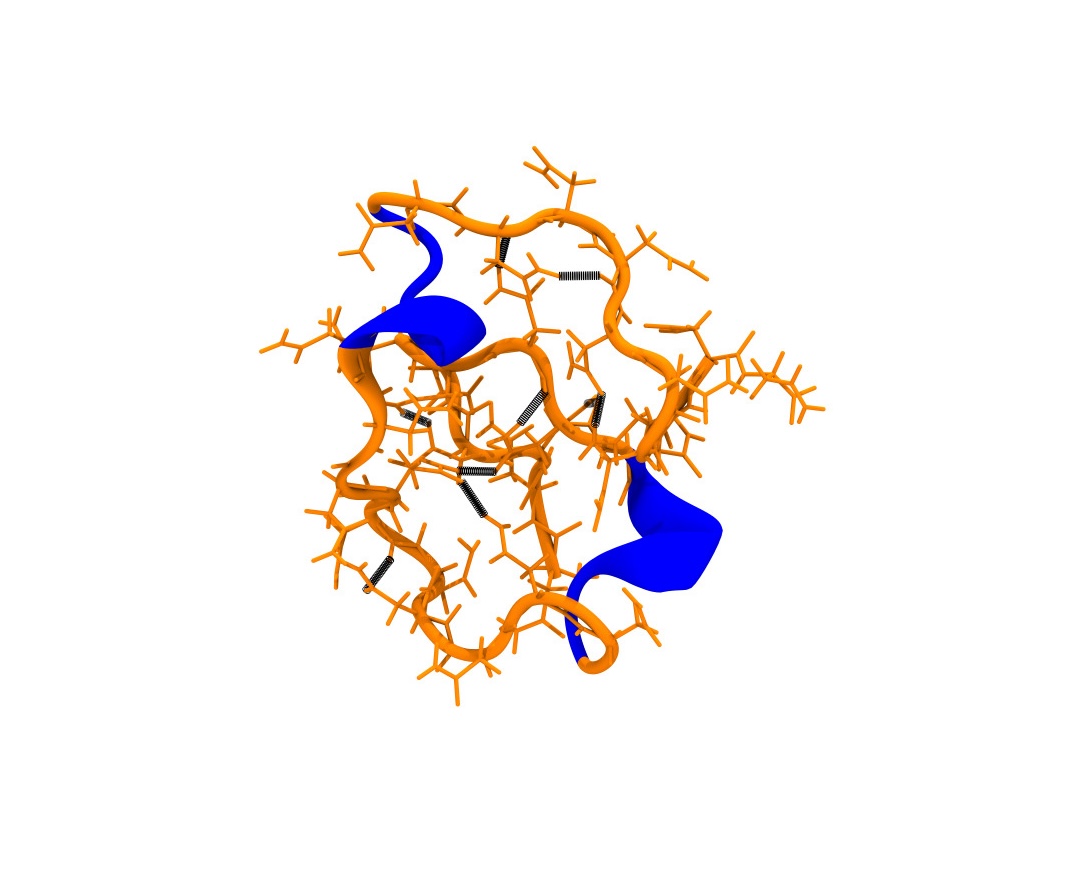
Hydrophilic core
The image depicts a synthetic peptide of 10 leucines with 30 asparagines. The dense amount of asparagines form many hydrogen bonds, creating a "hydrophilic core". In this case, these weak interactions are the dominant interaction that stabilize the peptide. Hydrogen bonds are represented as black springs.
Image Credit: Ryan Lamb
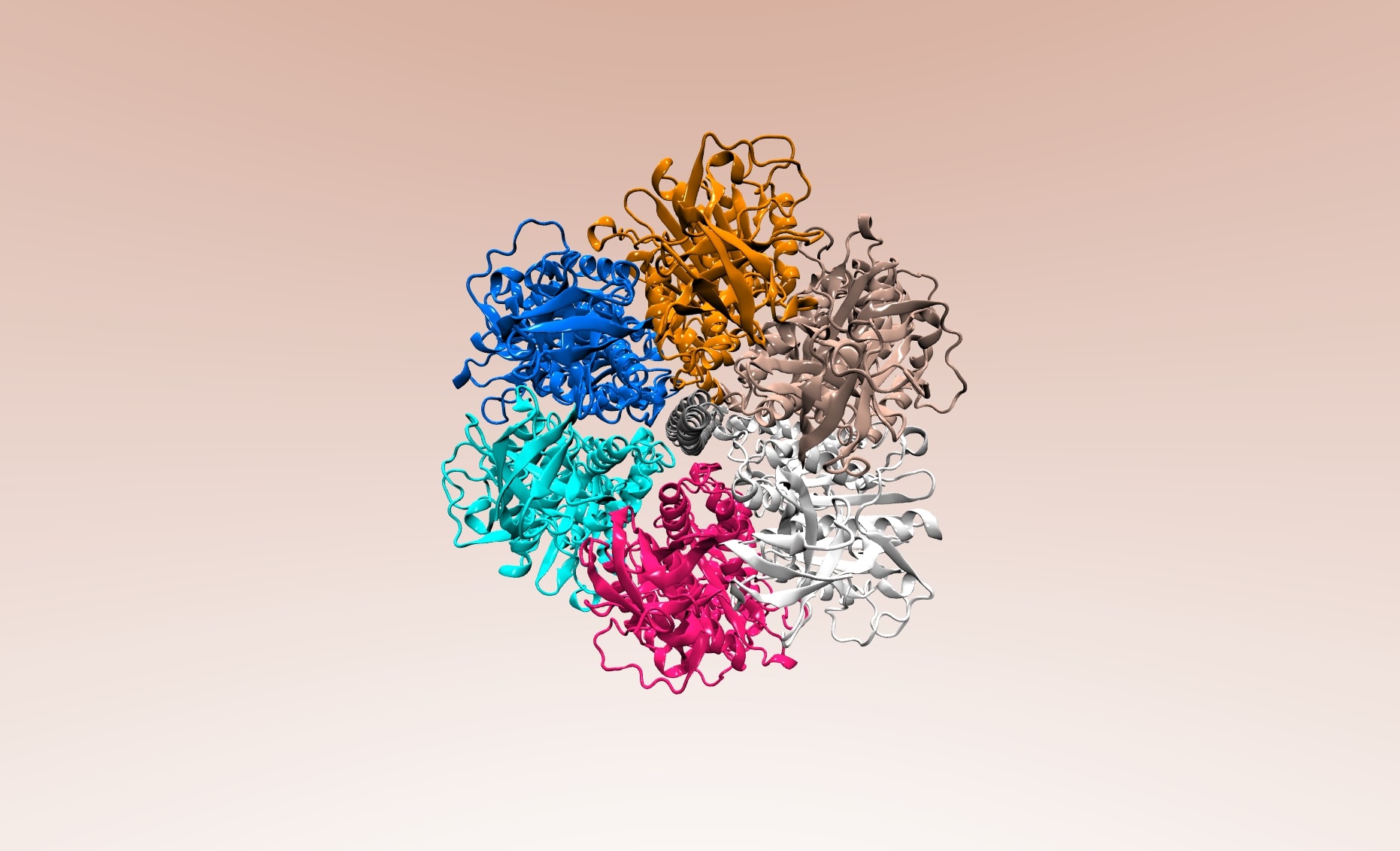
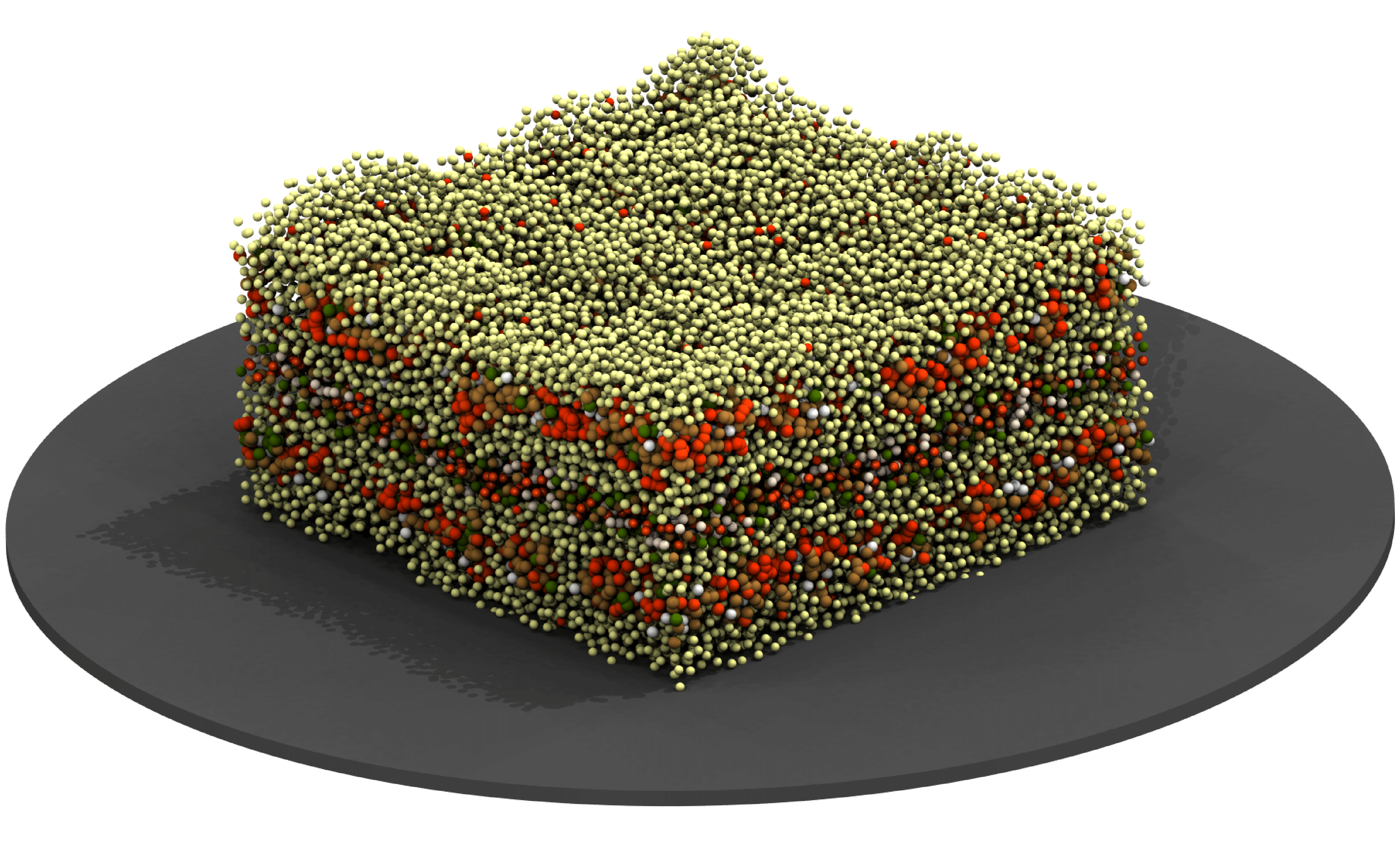
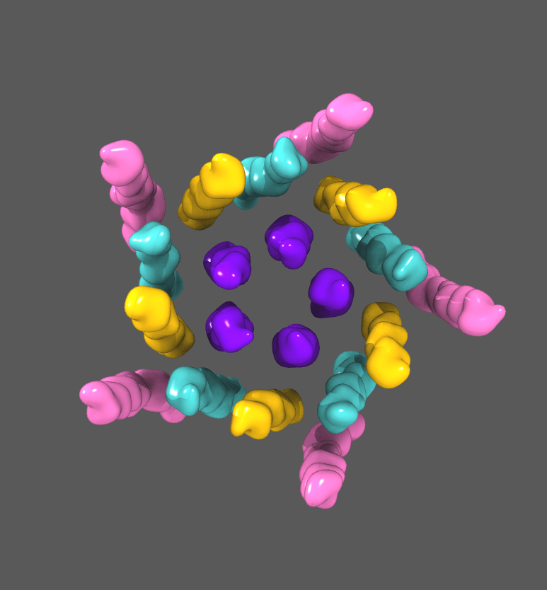
Structure in Sync
A structure of bovine mitochondrial F1-ATPASE (PDB: 1BMF). Each polypeptide chain is shown in a distinct color to present a symmetrical quaternary characteristic.
VMD ENTRY BY A BEGINNER CONTEST WINNER 2025
Image Credit: Asim Dave
Bubbles
Insulin, with amino acids colored by their predicted sensitivity to mutation. Mutations to red residues are likely to be pathogenic, while mutations to blue residues are likely to be benign. Grey residues had no prediction available. Mixed coloring indicates a mix of predicted effects. Predictions made using EVE (https://evemodel.org/).
VMD IMAGE CONTEST WINNER 2024
Image Credit: Connor Pitman
Crystal Structure
Lipids are extremely flexible small molecules. This is made more obvious as the lipid is phased out of a system during a free energy perturbation simulation. In these simulations, they system starts in a "normal" state - all the atoms are interacting with all the other atoms. But as the simulation progresses, the interactions between the lipid of interest (here POPG) and the rest of the simulation are turned off. The relative rigidity of the protein (ELIC) is emphasized by being represented as a crystal-like surface (blue). This is contrasted with the molten, liquid-like appearance of the POPG molecule (brass). The transparency of the lipid approximately corresponds to the strength of its interactions with the rest of the simulation system. Water and other lipids not shown.
VMD MOVIE CONTEST WINNER 2024
Movie Credit: Ezry Santiago-McRae
Lasagna
Similar to lasagna, cell membranes are layered structures. Just as the pasta layers in lasagna provide essential support for the meat, cheese, and sauce, the cell membrane offers structural support crucial for the cell's survival. Without a membrane, a cell would resemble a lasagna without pasta, a mess.
VMD IMAGE CONTEST RUNNER UP 2024
Image Credit: Alejandro Dagnino
Like a fish
in water
Unlike folded proteins that have specific native states, IDPs are unstable and fluctuate in space, twisting and turning, stretching and shrinking. BDNF (orange) is shown to exist in an ensemble of conformations, vibrating at different speeds over a short period of time. It moves in a staccato manner similar to a small orange fish swimming through the waters of a fast-paced river.
VMD MOVIE CONTEST RUNNER UP 2024
Movie Credit: Lindsey Riggs
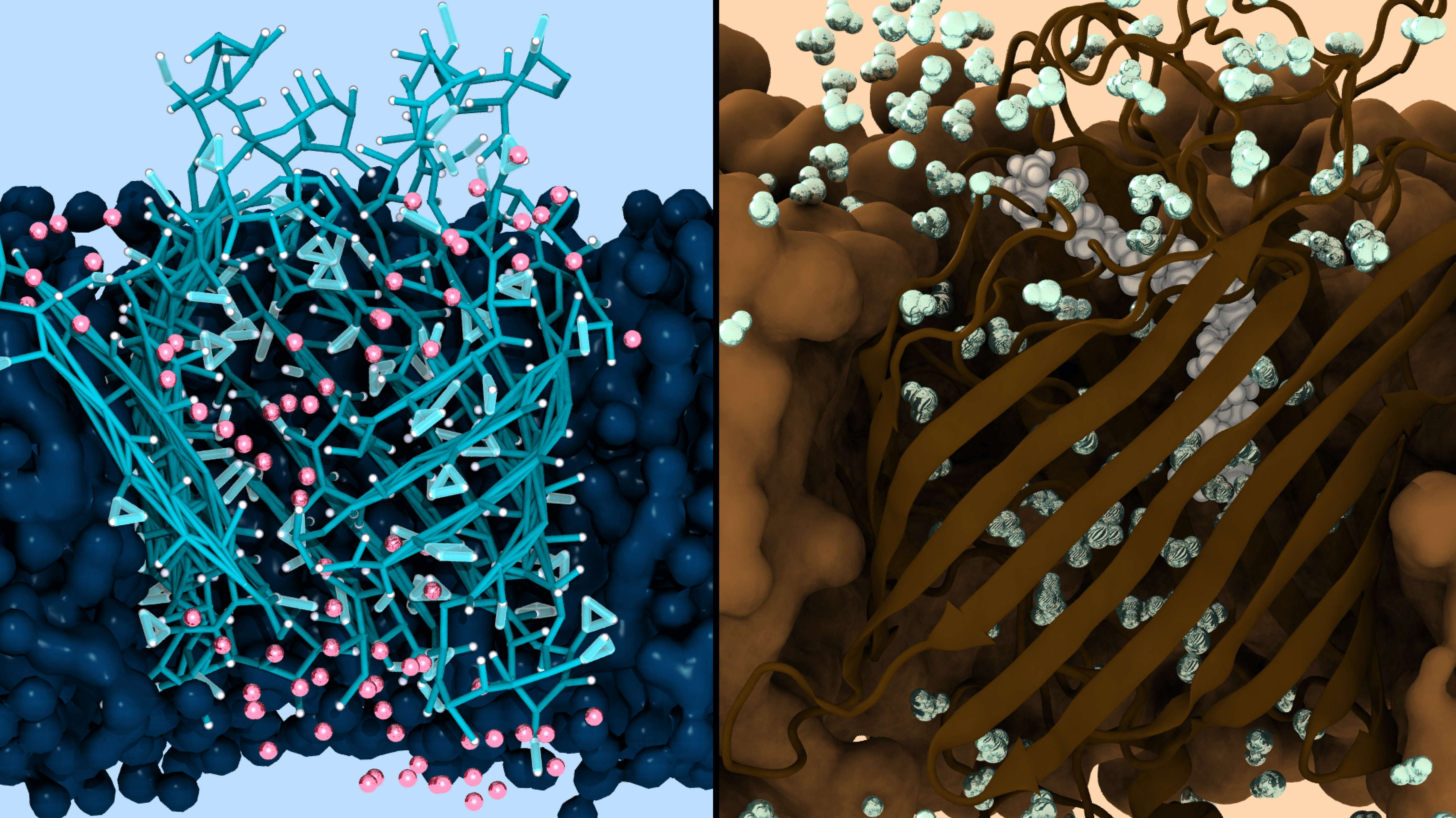
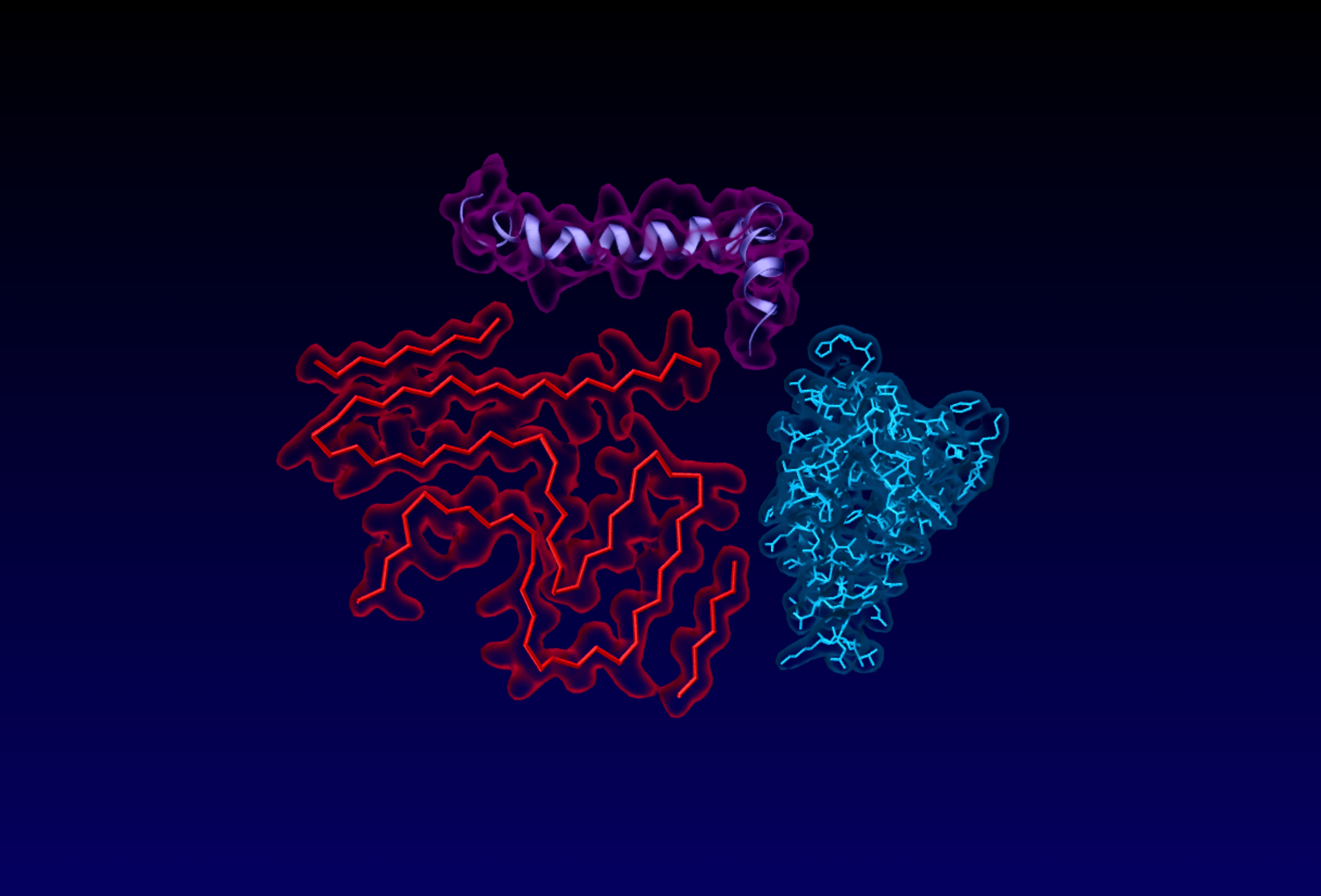
Duality
Left: This image is from a coarse-grained simulation of the open structure of BamA, the beta-barrel and surrounding lipids are in blue. Water within 8 angstroms of the protein is shown in red. This coarse-grained structure can be backmapped to atomistic resolution.
Right: This image is from an atomistic simulation of the closed structure of BamA, the beta-barrel and surrounding lipids are in brown. There is a lipid trapped in the center of the beta-barrel shown in white. Water within 10 angstroms of the protein is shown in blue. This atomistic structure is used as the starting point for coarse-graining.
Image Credit: Jahmal Ennis
Visualizing
Hydrophobicity
5j8v the breaker of programs, blobulated by the VMD blobulator GUI. Without the GUI, using just the blobulate proc feature, this would take 20 to 30 minutes of entering graphical representation settings in VMD.
Image Credit: Ryan Lamb
Burger King
The transmembrane domain of the nicotinic acetylcholine receptor, as it would have been decorated if it were a Burger King in the 90s.
Image Credit: Jesse Sandberg
Fuzzy Functions
Amyloid beta (purple), alpha-synuclein (red), and huntingtin (blue) are well-known IDPs involved in the development of Alzheimer’s, Parkinson’s, and Huntington’s disease respectively and are shown in their awareness colors. Uncovering the “fuzziness” of an IDP may spread light on its function, possibly paving the road to therapeutics for these neurodegenerative diseases.
Image Credit: Lindsey Riggs
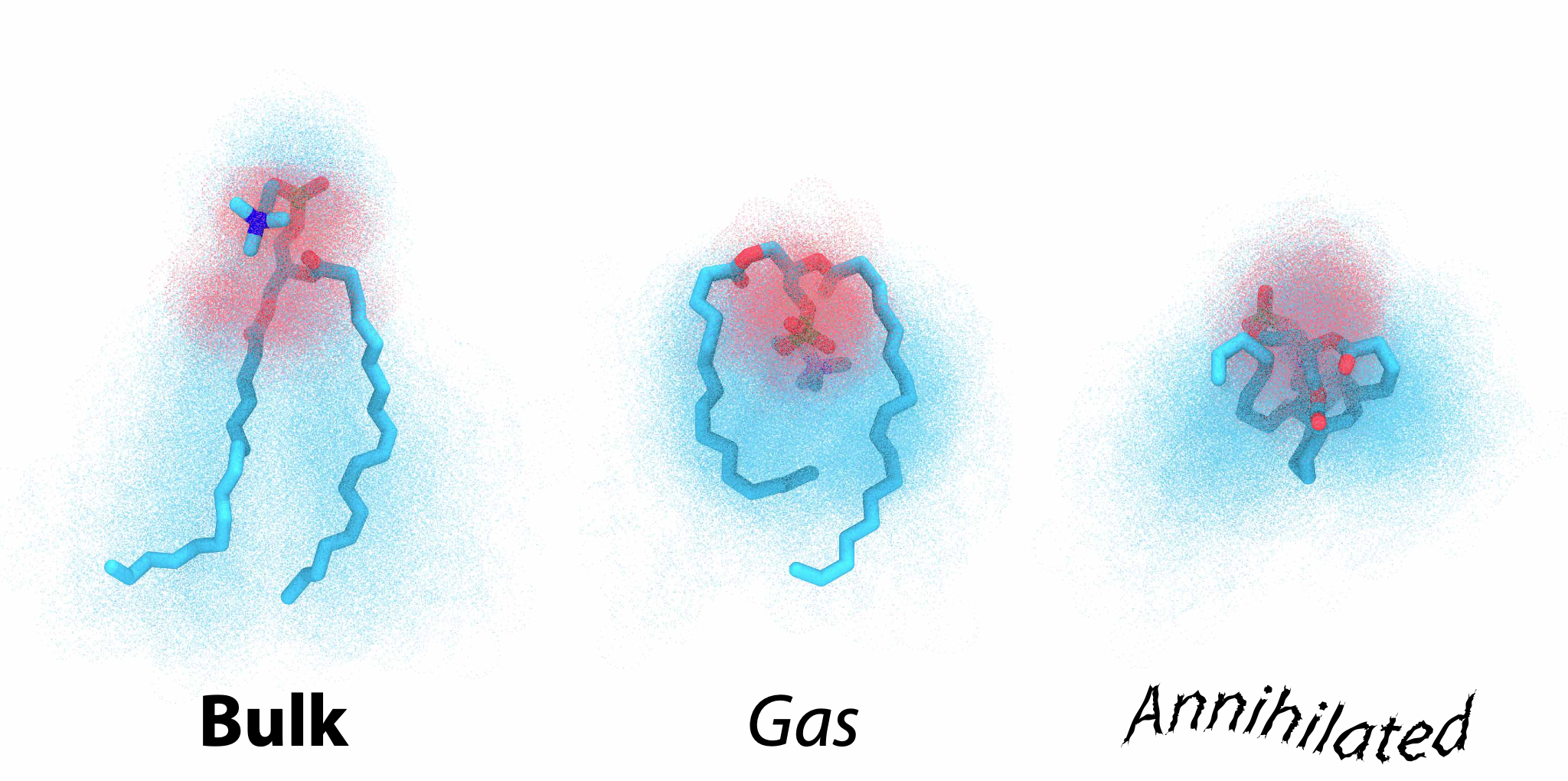
Three Faces
of POPC
In each image the accessible conformations are represented as a cloud around a representative pose from the respective phase. The three phases are (left to right) bulk POPC membrane, gas, and annihilated. The annihilated phase is like gas phase but all non-bonded interactions have been turned off allowing the lipid to pass through itself.
VMD IMAGE CONTEST WINNER 2023
Image Credit: Ezry Santiago-McRae
Tinker Toys and
Magnetic Balls
A POPC Membrane with two 2 nm gold nanoparticles embedded in it. We observe two polar nanoparticle covered in hydrophobic ligands aggregating. Nanoparticles cause unfavorable membrane conformations due to the exposed regions interacting with water. The membrane rearranges aggregating nanoparticles with ligands splayed open, alleviating unfavorable lipid configurations.
VMD MOVIE CONTEST WINNER 2023
Movie Credit: Jahmal Ennis
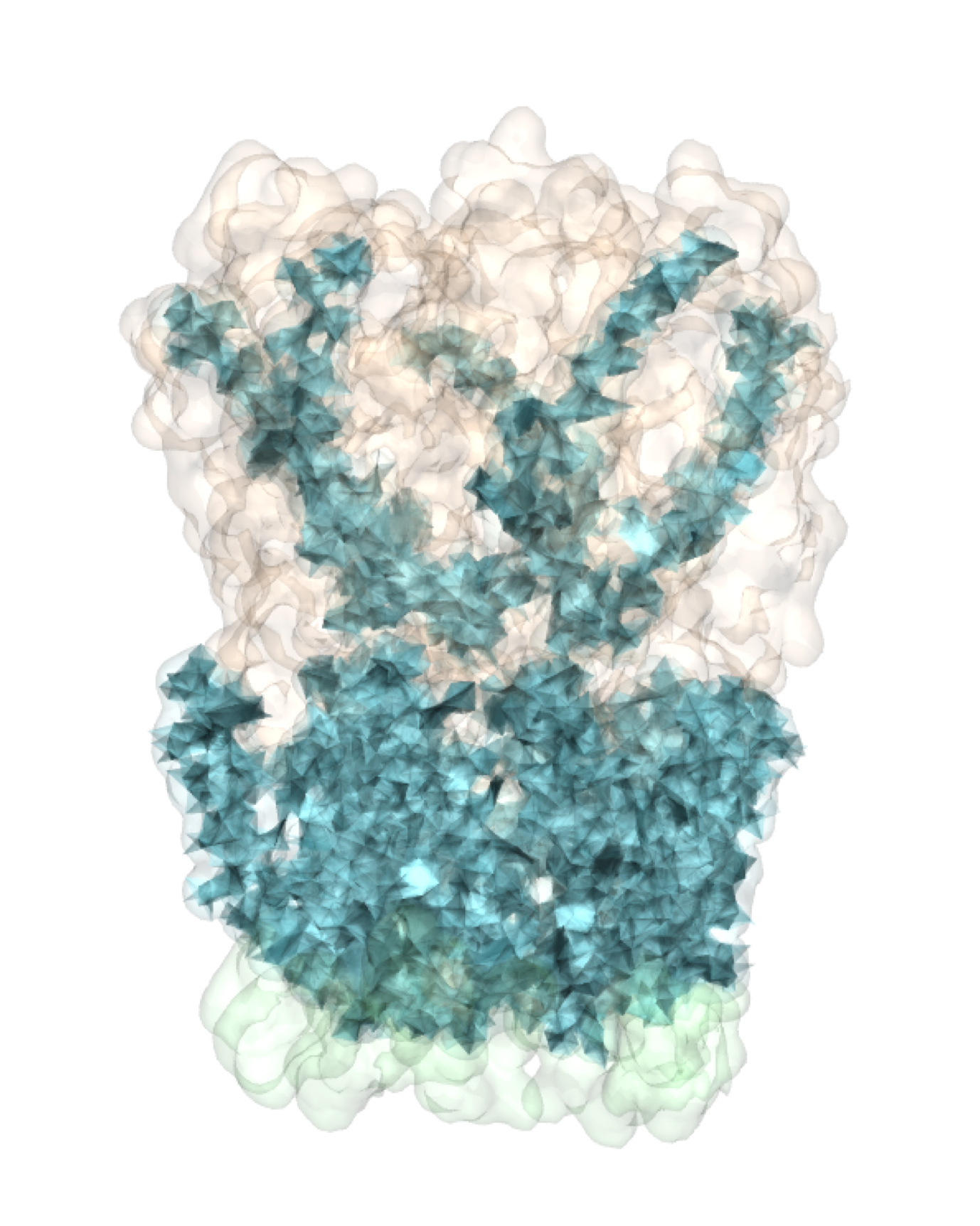
Crystalized!
Wildtype ELIC colored by contiguous hydrophobicity (blobs), defined by the blobulator. Blob type assignment is a result of blobulation settings chosen to detect transmembrane domains. p- (polar) and s- (short) blobs are colored orange and green, respectively, h-blobs (hydrophobic) are slightly opaque and colored blue.
VMD IMAGE CONTEST RUNNER UP 2023
Image Credit: Connor Pitman
Frame of Reference
The video shows two perspectives on decoupling. The left panel shows the view of the protein/environment while the right panel shows the view of the lipid. The audio is the raw value of the delta E (the difference in energy between adjacent lambda windows); higher volume corresponds to larger values of delta E. The first half of the video is quite tame because only electrostatics are being decoupled - the environment and the lipid still “see” each other for the most part. The Van der Waals interactions are then scaled out over the second half of the video denoted by both a change in volume and the visual fading of either the lipid (left) or environment (right). The last moments of the audio have a rapid increase in volume as we approach the discontinuity between slightly coupled and fully decoupled.
VMD MOVIE CONTEST RUNNER UP 2023
Movie Credit: Ezry Santiago-McRae
For the Love of
Hydrophobicity
A POPC Membrane, both waters and ions hidden.
Image Credit: Jahmal Ennis
Ribbons
The Envelope (E) protein of SARS-CoV-2 (grey) embedded in a thick, poly-unsaturated membrane (magenta). Only the hydrophobic core of the membrane is shown.
Image Credit: Jesse Sandberg
Landscapes of
Alien Nature
The image shows 5 nanometer gold nanoparticles
embedded in a POPC membrane. The POPC headgroups are shown in a iceblue, tails are in purple, GNP’s are shown in cyan, and ligands are hidden. These GNP’s bend the membrane into various shapes leading to areas of high curvature.
VMD CONTEST WINNER 2022
Image Credit: Jahmal Ennis
Envelope protein of
SARS-CoV-2 wearing a ruffled skirt
The pleats of the skirt represent the trajectory-averaged height of the outer membrane C1A/B beads as a function of r and theta. Coloring indicates mean curvature of the membrane surface. Positive curvature is shown in blue; and negative curvature is shown in red.
Image Credit: Jesse Sandberg
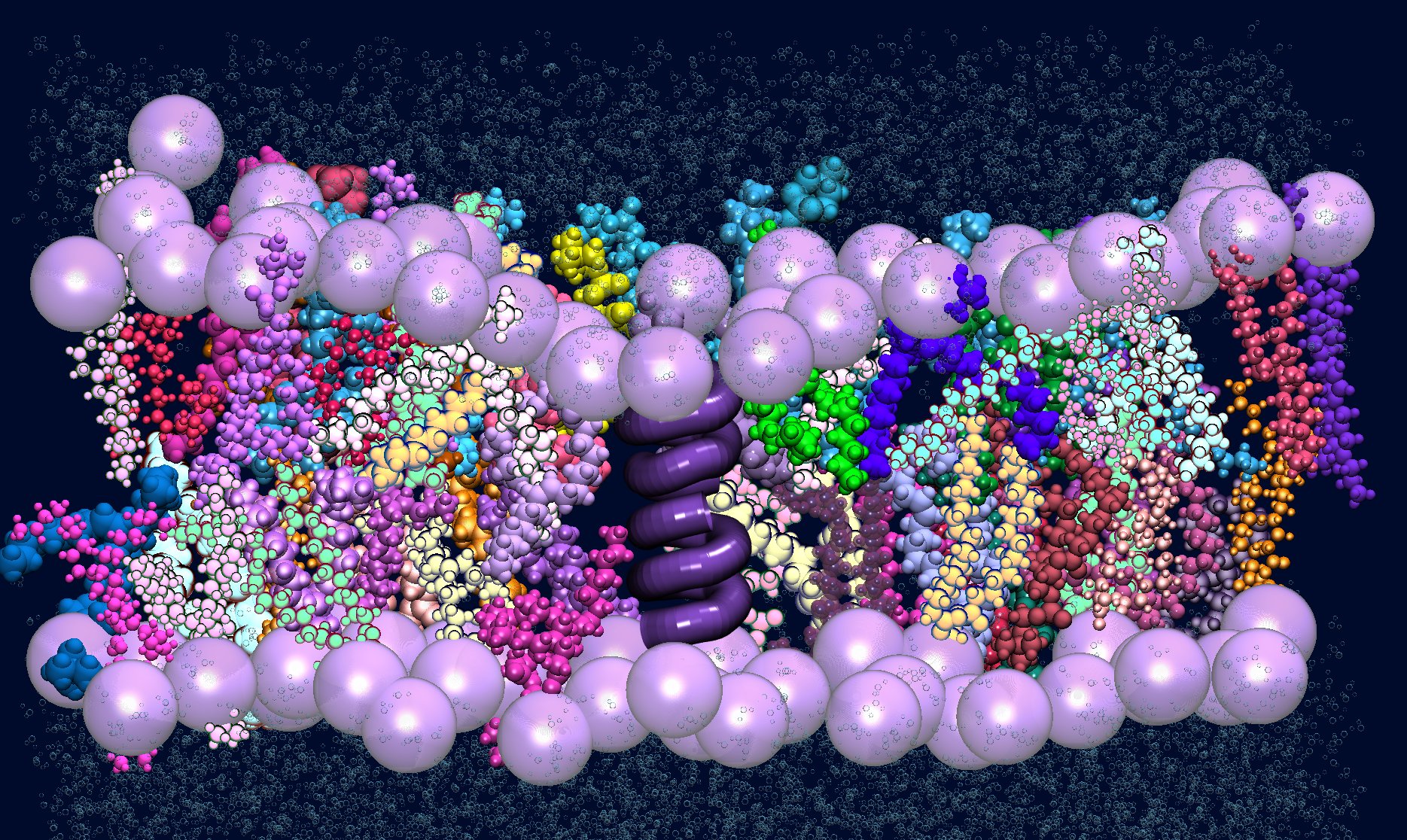
The most colorful lipid bilayer membrane!
The lilac beads represent the phosphate head groups. The multicolor chains represent POPC and the purple-ish secondary structure protein is Gramicidin A (warning: Gramicidin A is not a protein, it is a peptide). The bubbles represent water beads.
VMD CONTEST WINNER 2022
Image Credit: Mariadelia Arguello Acuna
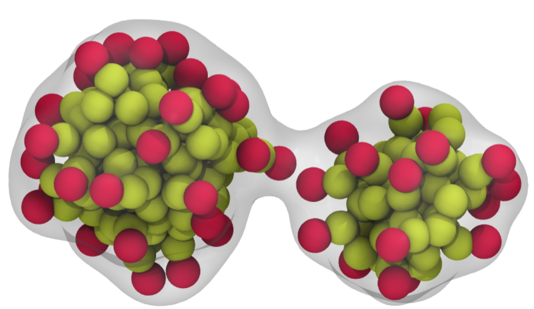
SDS Micelles getting
to know each other
SDS micelles coming into contact just before fusing together! The negatively charged headgroups are shown in red, and hydrophobic tails are shown in yellow.
Image Credit: Connor Pitman
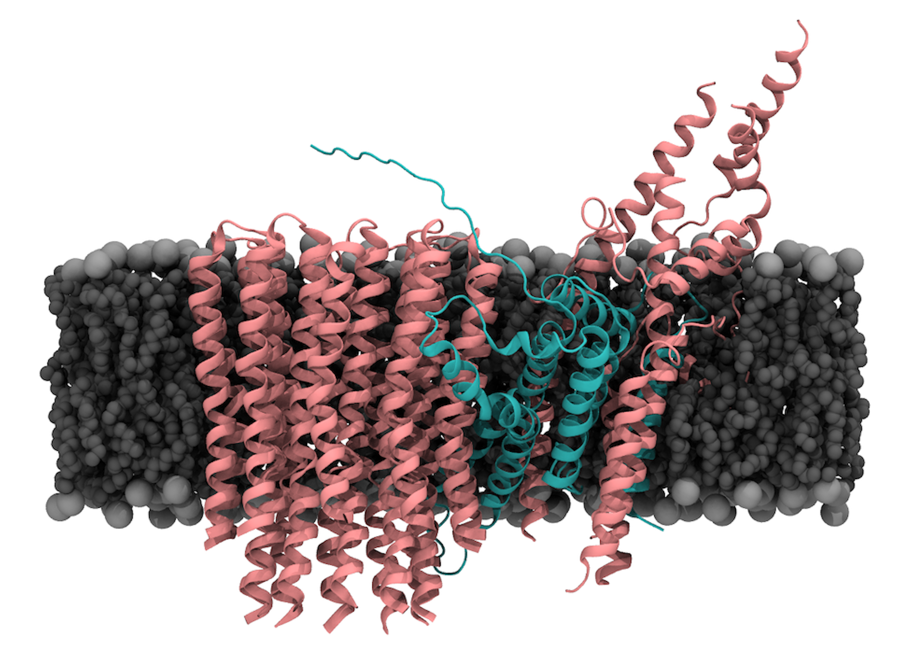
A Protein Subunit Gone Wrong
The F0F1 ATP Synthase is a highly conserved mitochondrial membrane protein, responsible for ATP synthesis. It synthesizes ATP using a proton gradient across the membrane. Ice worms have a unique Histidine-rich extension in the APT6 subunit (shown in cyan) that is hypothesized to accelerate proton flow across the membrane, thus producing more ATP levels than the energetic demand of ice worms. Shown in pink and cyan is the predicted structure of the ice worm F0 domain embedded in a POPC bilayer.
Image Credit: Noureen Abdelrahman
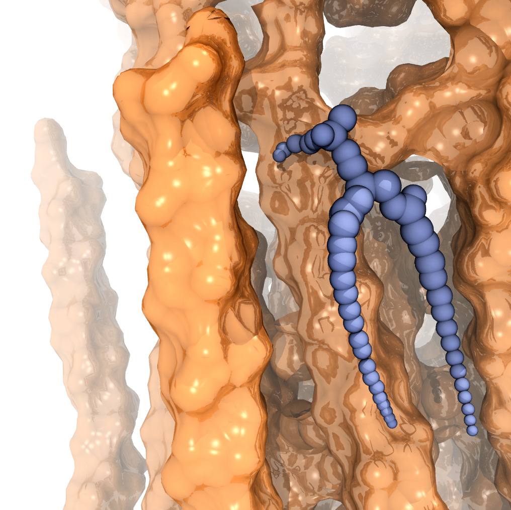
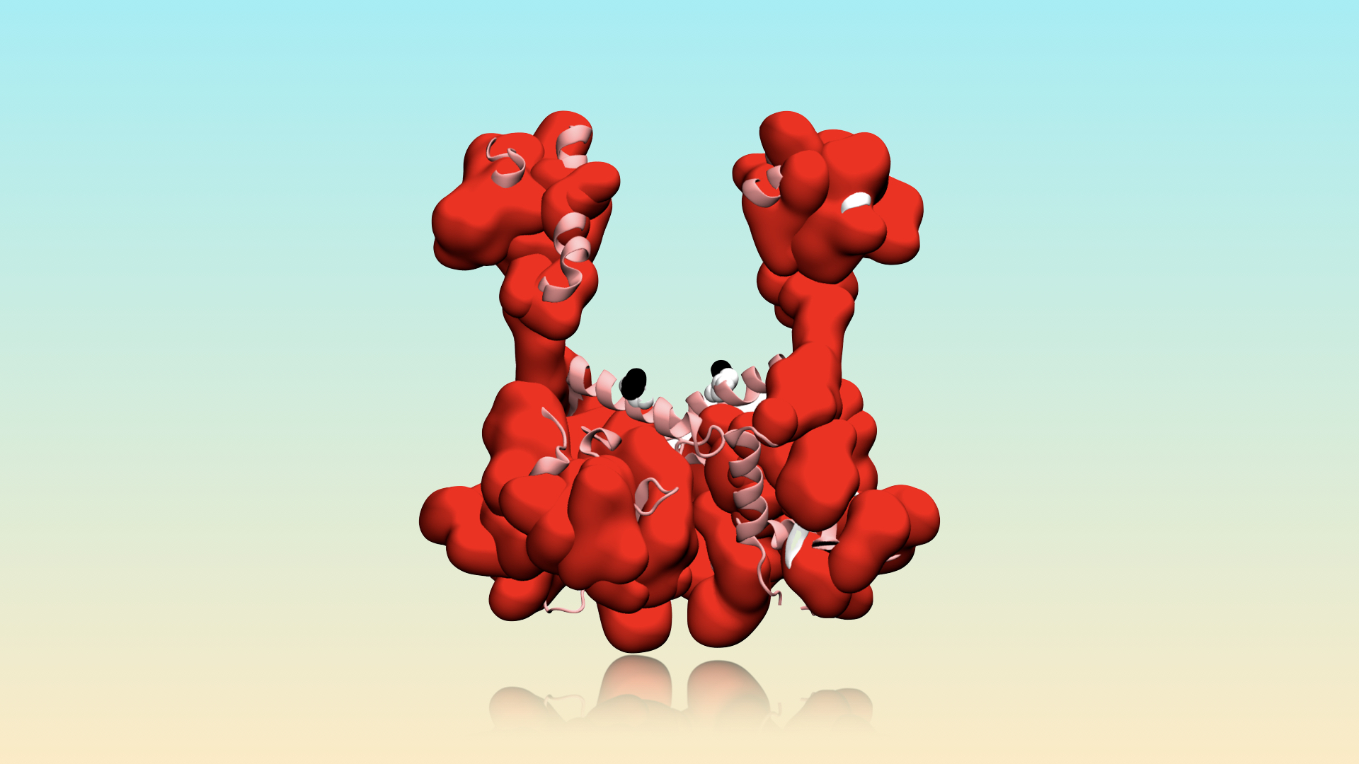
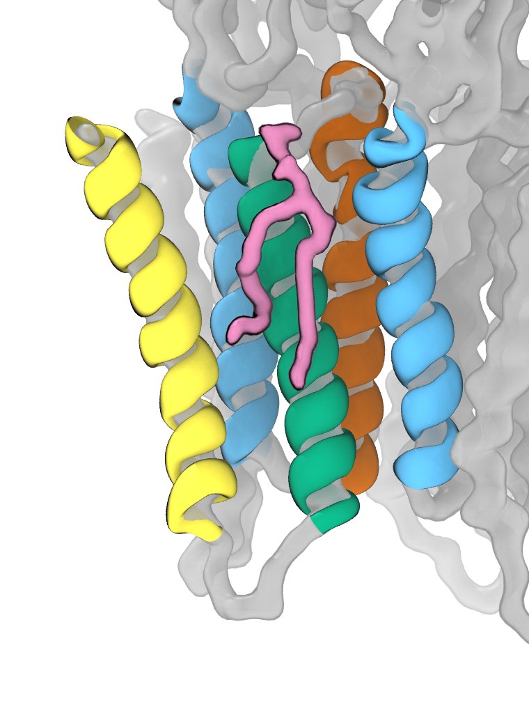
Membrane Lipid in a Pocket
The Erwinia ligand-gated ion channel (ELIC) is a bacterial homolog and model system for important neuronal membrane proteins. Ligand-gated ion channels typically only open after binding a ligand. Membrane lipids, however, can have a tremendous impact of the function of these membrane proteins. POPG is shown in pink; transmembrane helix 1 (M1) in blue; M2 in orange; M3 in green; M4 in yellow. Portions of the protein not immediately relevant to the binding pocket are shown in gray.